Monday, 19 November 2007

TRANSFORMATION OF E.COLI WITH PLASMID DNA

3. TRANSFORMATION OF E.COLI WITH PLASMID DNA – Dr. Fukada

  • Gene(s) of interest(s) can be isolated from organisms such as plants, bacteria or animals, and introduced to bacteria preferably E. coli for amplification. This is known as cloning. Thereafter, one can modify or analyze the gene and then insert it back to the organism. E. coli has the ability to take exogenous DNA naturally.
  • Two methods are used to transform E. coli namely:

1. Chemical transformation

a) Hanahan method - which uses calcium ions. The transformation efficiency of this method is 5 x 10⁷ – 1 x 10⁸ / µg of DNA, hence is unstable.

b) The Cacl2 method – has a lower transformation efficiency of 5 x 10⁶ to 2 x 10⁷.

2. Physical transformation – mainly electroporation. This method uses high electric voltage to introduce the DNA into bacteria. The transformation efficiency of this method is 1 x 10⁸ to 1 x 10¹⁰

  • Two plasmids A and B, with sizes 3.5 kbp and 11 kbp respectively were used to transform E. coli and the results were analyzed and compared. The reagents and media for use and the electrocompetent cells were prepared.
  • It was noted that 3 factors affected the transformation efficiency. These are:

Optical Density (OD) of bacterial culture that is usually 0.4 to 0.6 for electroporation, and it takes about 2-3 hours to attain this OD.

Temperature – all processes must be done at low temperatures or in ice, it is necessary to keep the bacteria alive. All reagents and instruments must be cooled before use.

Purity of the reagents – this is important for chemical transformation methods. Both water and reagents to be used must be pure. The supernatant must be completely removed before addition of the next solution. After centrifugation, the tube must be inserted into ice in slanting position and the surplus supernatant removed completely.

· For both plasmids, 2 µl of each plasmid was separately added into 100 µl of E. coli culture LB medium, mixed well, and the whole amount transferred to electroporation cuvette. 500 µl of LB was subsequently added to give a total volume of 600 µl and pulsed. 100 µl of mixture was picked and added into 900 µl of LB to obtain a diluted culture, while the remainder (500 µl) was undiluted.

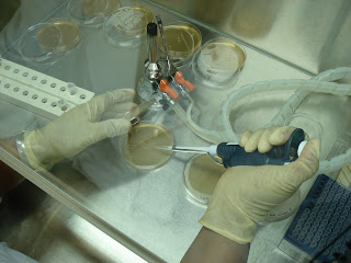
· For both suspensions, (diluted and undiluted), 100 µl was put on LB agar plates and the suspension spread across the plates by rolling sterile glass beads. The plates were incubated at 370 C overnight.

· The transformation efficiency of E. coli was expressed as the number of colonies per µg of DNA. Upon calculation, the results were displayed in the following table:

DNA

UNDILUTED

DILUTED

Plasmid A (3.5 kbp)

2.18 x 10⁸

2.31 x 10⁸

Plasmid B (11 kbp)

1.17 x 10⁷

1.5 x 10⁷

It was noted that the transformation efficiency was high in plasmid A. The ratio of transformation A:B was 18.6:1 (transformation efficiency of plasmid A was 18.6 times higher than that of plasmid B). This experiment demonstrated that small-sized plasmids have higher transformation efficiency as compared to larger-sized plasmids.

COMMONLY USED TECHNIQUES IN MOLECULAR CLONING

2. COMMONLY USED TECHNIQUES IN MOLECULAR CLONING – Dr. Sumitani

Preparation of reagents culture E. coli for preparation of chromosomal DNA

Media and reagents for growing E. coli were prepared. These included 200 ml Luria Bertani (LB) medium; Solution I (50mM Glucose, 25mM Tris.Hcl, 10 mM Ethylene Diamine Tetra Acetate - EDTA); Solution II (0.4N NaOH and 0.2% SDS); Solution III (3 M potassium acetate and 2 M acetic acid) were prepared by the participants in different groups. EDTA is a trap that inhibits enzymatic reaction. All these were autoclaved with the LB medium divided into test tubes. The media was allowed to cool and inoculated with E. coli aseptically in the fume hood and subsequently incubated at optimum conditions to allow for growth.

Genomic DNA from E. Coli

DNA is purified by phenol extraction. This removes protein and lipid contaminants. The DNA is neutralized with buffer and then mixed with chloroform and isoamyl-alcohol. 8-quinolinol and 2-mercaptoethanol were subsequently added.

It is noteworthy that phenol is easily oxidized and has an aqueous phase to prevent oxidation thus acts as a buffer.

Alcohol precipitation: 2-3 volumes of Ethanol are required for DNA precipitation and 2-propanol (0.6 to 1 volume of sample).

Salts required: 3 M Na-acetate for 1/10 volume of sample and 5 M NaCl for 1/20 to 1/30 volume of sample. NaCl is recommended for use with samples containing SDS as it gives better results.

Preparation of chromosomal DNA

Obtaining chromosomal DNA is procedural as follows:

  1. Break down the cell by Proteinase K and SDS. Proteinase K is a non-specific proteinase that acts on the surface of the cell membrane while SDS is a surfactant that breaks the plasma membrane.
  2. Removal of macromolecule contaminants (proteins, lipids and polysaccharides). Many compounds are contained in the cell and it is therefore advisable to remove the macromolecules first because they concentrate. The Cetyl tri-methyl ammonium bromide (CTAB) method is used to remove polysaccharides. CTAB is a cationic surfactant and it can easily complex with polysaccharides and precipitates.
  3. Nucleic acids (DNA and RNA) are concentrated or precipitated by 2-propanol and NaCl (used because it is SDS hence used for better results).

The cells were collected by centrifugation of bacterial culture at 10 000 rpm for 1 minute at 40 C and the supernatant completely removed by pipetting. 567 µl TE buffer was added to suspend the cell wall and 30 µl of 10% SDS and 3µl of 20 mg/ml proteinase K were added respectively, thoroughly mixed and incubated at 370 C for an hour. Thereafter, 100 µl 5 M NaCl was added and mixed thoroughly, and 100 µl CTAB/NaCl solution added, thoroughly mixed and incubated for 10 minutes at 650 C. An approximate volume (0.7 to 0.8 ml) of chloroform/iso-amylalcohol was added, thoroughly mixed and spinned at 15 000 rpm for 5 minutes at 40 C. The aqueous phase was transferred to a new tube and treated with 0.7 ml phenol/chloroform/iso amylalcohol in a ratio of 25:24:1. This was mixed thoroughly and spinned at 15 000 rpm for 5 minutes. The aqueous phase was again transferred to a new tube and 0.4 to 0.5 ml of 2-propanol added and mixed well. By this time, the DNA precipitate becomes visible (clear). The DNA precipitated was transferred to a new tube and 1 ml of 70% Ethanol was added, centrifuged at 15 000 rpm for 5 minutes at 40 C. The supernatant was completely removed and DNA pellet was allowed to dry up by leaving the lid open for 20 to 30 minutes. The DNA was dissolved in 100 µl TE Buffer, 1 µl of 20 mg/ml RNase A was added, mixed and stored in the refrigerator.

Preparation of plasmid DNA from E. coli

Plasmid is not required for E. coli to grow. Plasmid DNA tends to discard from the cell. To maintain plasmid DNA in the cell, antibiotics are added into the media. Two plasmids were used, pUC18, with 100-200 copies per cell and pBR322 which as 30-50 copies per cell. The difference between the plasmid Puc18 and Pbr322.

Extraction and purification of plasmid DNA

Chromosomal DNA has several mega bases while plasmid DNA has several thousand bases. In bacteria, the plasmid does not appear in circular shape. The plasmid DNA was to be separated from the chromosomal DNA that was previously obtained. In alkaline conditions, double stranded DNA dissociates to single stranded DNA as the hydrogen bonds break down. Plasmid DNA that is in straight form does not fully dissociate. The DNA solution was subjected to alkalinic conditions then was rapidly subjected to neutral conditions. The plasmid DNA easily re-anneals, unlike the chromosomal DNA which is large.

Procedure:

  1. The bacterial cells were broken down by SDS, and Sodium hydroxide.
  2. Plasmid DNA was separated from the chromosomal DNA by neutralization by potassium acetate.
  3. Proteins were removed via phenol extraction, as the resultant solution contained plasmid DNA and RNAs.
  4. Salts and low molecular weight compounds were removed by Ethanol precipitation
  5. RNAs were removed by RnaseA.

Preparation of Agarose gel for Electrophoresis

1.0 g Agarose

100 ml 1 x TAE buffer

Agarose solution was melted in the microwave until it bubbled, poured into the gel tray and solidified by cooling.

The obtained DNA (10 µl) and 2 µl loading buffer was put into the wells of the gel by pipetting, with lambda DNA/Hind III marker.

PREPARATION FOR LABORATORY WORK

1. PREPARATION FOR LABORATORY WORK - Iwaki Sensei

The laboratory where we carried out all the practicals and lectures for the training is physically protected P1 level laboratory. No foods and drinks are permitted in this particular lab. The hoods are fitted with High Efficiency Particulate Air (HEPA) filters and safety cabinets.

Garbage disposals are in various categories namely:

1. Combustibles e.g. papers, paper towels

2. Non-combustibles e.g. gloves, plastic tips and tubes

3. Glassware waste buckets for broken glassware and cleaned with broom and collection tray

4. Biological waste such as petri dishes and cells, are to be put in autoclave bags for autoclaving before disposal.

5. Sharp objects such as blades, needles and syringes are disposed off separately.

It was well noted that the university has 4 kinds of garbage waste bins namely:

1. Cans

2. Pet bottles

3. Papers

4. Plastics

All glassware should be washed well with detergent and final rinsed with de-ionized water. The room should be kept clean as it is also for biochemical experiments besides the lectures. The supervisor designated for that day oversees that all the above is observed by the participants.